Choosing the right Computerized Foot Scanner For Orthotics is a clinical and operational decision. It affects assessment quality, fabrication accuracy, patient communication, and daily workflow. The global orthotics market continues to expand as clinics adopt digital measurement and customized devices. Grand View Research’s 2024 foot orthotic insoles market analysis identifies customization and digital technologies as important growth drivers. However, market growth does not guarantee clinical value.
A reliable scanner should capture foot length, width, arch height, heel position, and relevant pressure patterns. It should also produce repeatable results under normal clinical conditions. The 2023 International Working Group on the Diabetic Foot guidelines emphasize structured foot assessment and pressure-related risk management. A scanner can support these tasks. It cannot replace vascular checks, neurological testing, gait observation, or professional judgment. That distinction matters.
Small details often reveal major differences. Can the system scan a standing foot without movement artifacts? Does it record left and right feet separately? Can clinicians export measurements into CAD or orthotic manufacturing software? Is calibration simple enough for a busy treatment room? These questions deserve practical testing, not attractive sales claims. A 2024 report from MarketsandMarkets also links digital healthcare adoption with demand for automated measurement tools, but commercial reports may simplify clinical realities. That is worth remembering.
This guide presents seven practical tips for evaluating scanner accuracy, software, compatibility, training, maintenance, data security, and total cost. Some recommendations may seem obvious. They are still frequently missed. The best device is not always the most advanced one. It is the system that produces dependable information, supports informed decisions, and fits real clinical habits. Expect trade-offs. Keep questioning them.
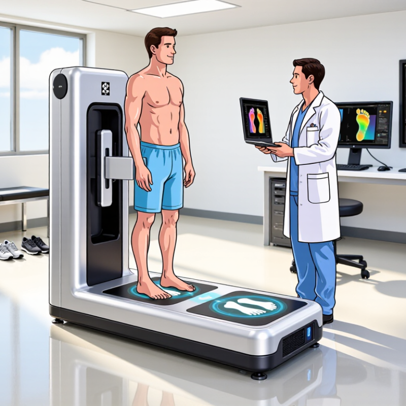
A computerized foot scanner should match the patients you actually treat. The IDF Diabetes Atlas, 10th edition, estimates 537 million adults had diabetes in 2021. That equals 10.5% of adults aged 20–79 worldwide. Many may need pressure-aware assessment, accommodative orthoses, or repeated fitting.
Define your clinical scope before comparing scanner features. For diabetic foot care, prioritize plantar-pressure capture, bilateral imaging, repeatable measurements, and simple data export. Check whether the system records arch height, foot length, heel width, and asymmetry. These details matter when swelling, neuropathy, or previous ulceration changes a patient’s loading pattern. The International Working Group on the Diabetic Foot recommends structured risk assessment and regular preventive care for at-risk patients.
Keep the workflow practical. A scanner should capture a stable stance, guide patient positioning, and produce files your clinical software can read. Ask for accuracy data, calibration procedures, cleaning instructions, and staff training requirements. Verify how measurements perform across different skin tones, mobility levels, and foot deformities. Real clinics are rarely perfect.
Do not treat a colorful 3D model as a diagnosis. It is an assessment aid. Pair digital findings with circulation checks, sensory testing, footwear review, and patient history. The IDF projects diabetes prevalence to reach 643 million adults by 2030, increasing the need for scalable clinical workflows. A less expensive scanner may seem attractive, but weak repeatability can create costly rework and misplaced confidence.
Patient needs should guide the purchase. Some patients require pressure assessment, accommodative orthoses, or repeated fitting. A scanner should match those cases.
Prioritize plantar pressure, bilateral images, arch height, foot length, heel width, and asymmetry. Swelling or neuropathy can change loading patterns. Measure both feet.
It should record a stable standing position and guide patient placement. The process should feel simple. Staff should not spend ten minutes correcting posture.
No. It is an assessment aid, not a diagnosis. Combine digital findings with circulation checks, sensory testing, footwear review, and medical history.
Open sample STL and OBJ files in the actual design software. Check millimeter units, mesh quality, surface normals, and file integrity. Look for gaps.
Import the same patient’s scans twice. Compare heel cup depth, forefoot width, and trim lines. Small alignment errors can cause uncomfortable orthotic changes.
Look for structured patient identifiers, secure exports, audit trails, and available healthcare data standards. Avoid closed portals that trap scan files. Access matters.
Use local patient numbers, scanning volume, remake costs, training, software fees, calibration, and staff time. Include referrals and wound-care expenses. The assumptions may be wrong.
Ask for accuracy results across skin tones, socks, weight-bearing positions, mobility levels, and repeated sessions. Request subject numbers and review methods. Perfect repeatability is unlikely.
Track calluses, skin changes, orthotic adjustments, referrals, and related care for six and twelve months. Review findings with a qualified clinician. A modest result can still matter.
Choosing a Computerized Foot Scanner For Orthotics requires more than comparing advertised features. Start by defining the clinical scope, especially when serving patients with diabetes; the IDF estimated that 537 million adults were living with diabetes in 2021. Evaluate whether the scanner’s accuracy, repeatability, and resolution are sufficient for consistent clinical decisions, rather than relying solely on sub-millimeter claims. Confirm that it can capture the foot under realistic weight-bearing conditions, supporting gait analysis, pressure assessment, and orthotic design.
Workflow compatibility is equally important. Check whether scans can be exported in common STL or OBJ formats and integrated smoothly with CAD/CAM systems and electronic health records. Finally, estimate the scanner’s return on investment by considering time savings, improved fitting, reduced remakes, and preventive care. This is particularly relevant because the ADA cites a 15–25% lifetime risk of diabetic ulcers. A well-matched scanner should improve clinical confidence while supporting efficient, measurable patient care.

 MedTrack